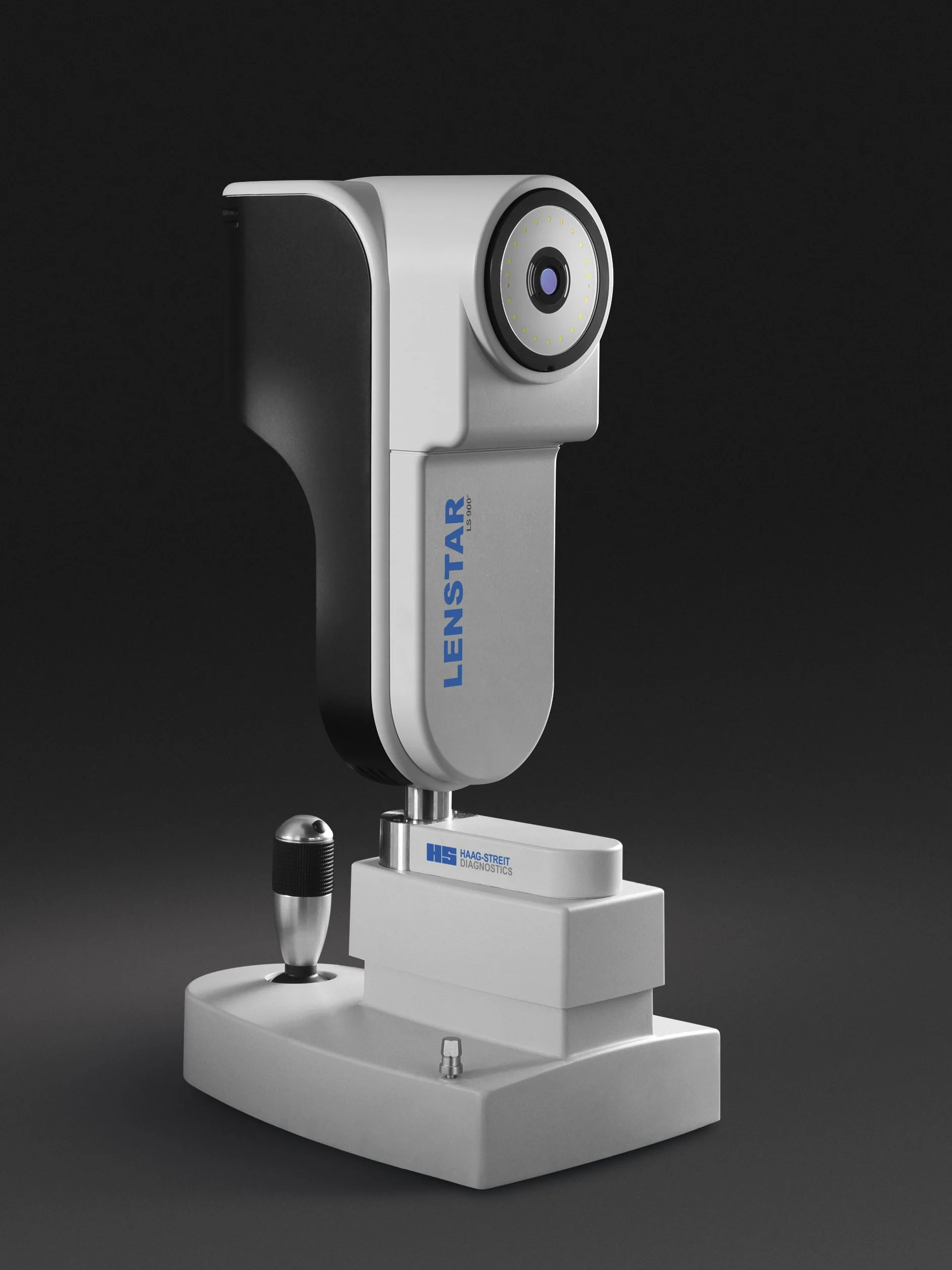
Lenstar Biometry for Myopia Management
The Haag-Streit Lenstar biometer measures the axial length of the eye, a critical measure for assessing the progression of myopia especially in children and adolescents
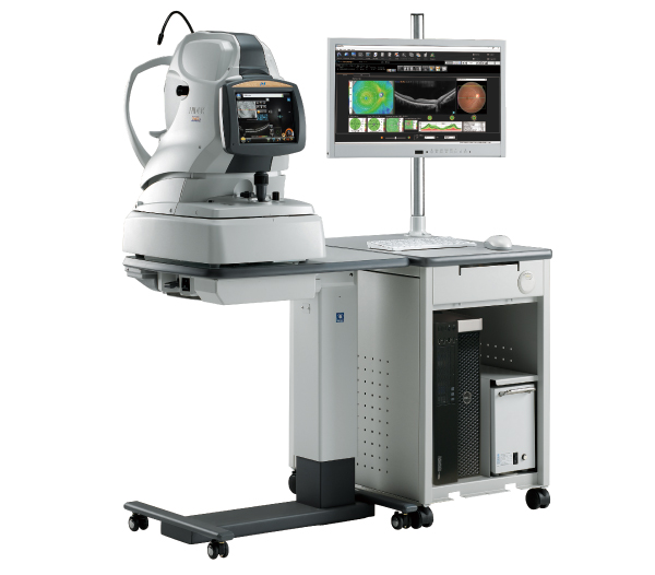
The Haag-Streit Lenstar biometer measures the axial length of the eye, a critical measure for assessing the progression of myopia especially in children and adolescents
Keratography with the Oculus K5 instrument is an invaluable tool for the assessment, diagnosis and classification of dry eye
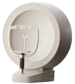
Measurement of visual fields by automated perimetry is an important technique for assessing glaucoma and neurological conditions
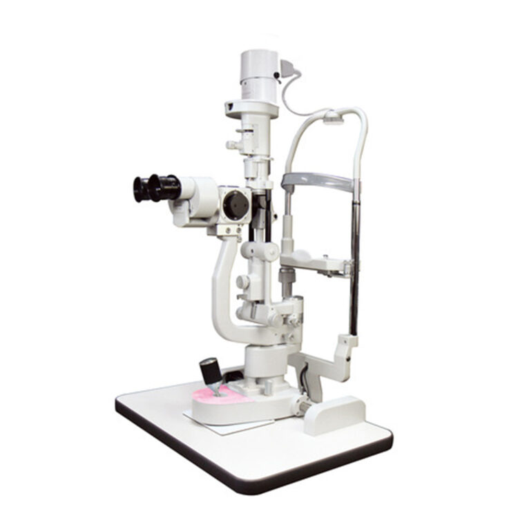
High quality magnification and lighting are essential for examining the front of the eye and detecting subtle changes, which is assisted further by the recording function of digital photography
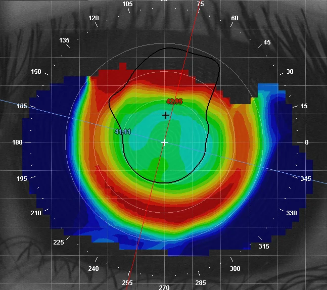
Corneal topography provides a computerised 3-D map of the front surface of the eye, with which we can see the characteristics of its curves and surface irregularities and which is enormously helpful for contact lens fitting
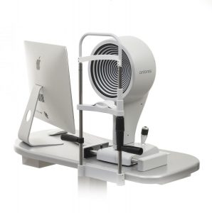
Our Optos Daytona scans the retina with two lasers through a range of 200°, where by comparison, standard digital retinal photography captures only the central 45°